
Figure 4.
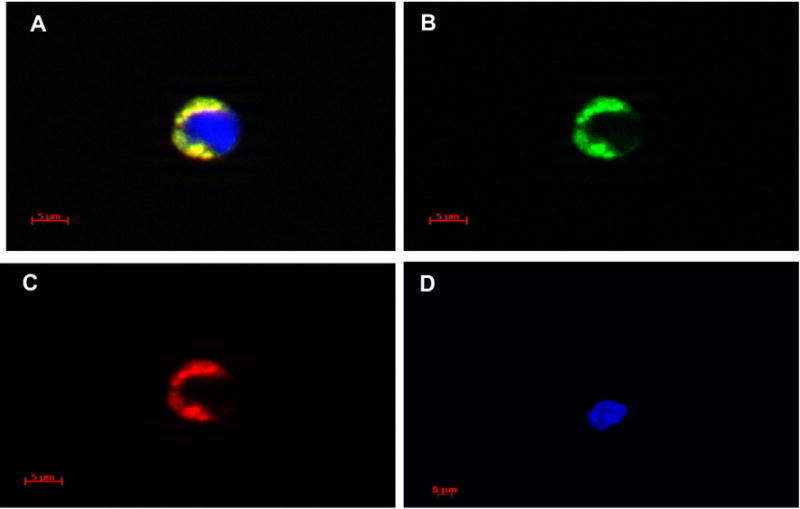
Digital images of bone marrow cells containing microvesicles. Examples of bone marrow cells containing PKH26 and CFSE labeled microvesicles are shown (A–C). (A) Composite image of one bone marrow cell taken through DAPI, FITC, and Texas Red filters. Blue color is DAPI (nuclear counter stain). (B, C) Images of the same cell taken through FITC and Texas Red filters, respectively. (D) Example of DAPI counterstained cell that has not taken up microvesicles image taken using DAPI, FITC, and Texas Red filters. Fluorescent images were taken with a Zeiss Axio Imager Z1 fluorescent microscope at 63 × magnification and Axiovision 4.6.3 software. Digital images were acquired using an AxioCam HRm. Three-dimensional images were taken utilizing a Zeiss ApoTome for structural imaging and a four-dimensional acquisition module.