
Figure 4.
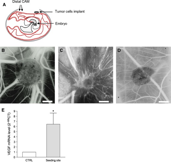
CAM assay. (A) Schematic representation of CAM assay. (B) Stereomicroscopic images of SK-LMS-1 embedded in Matrigel implanted on the CAM. After 4 days numerous allantoic vessels formed radially towards the implant. (C) 200 ng of human FGF-2 resospended in Matrigel was used as positive control. (D) Matrigel alone was used as negative control; scale bar: 1000 μm. (E) Quantification of avian VEGF in tumour-treated CAM samples by RT-PCR. *P < 0.05.