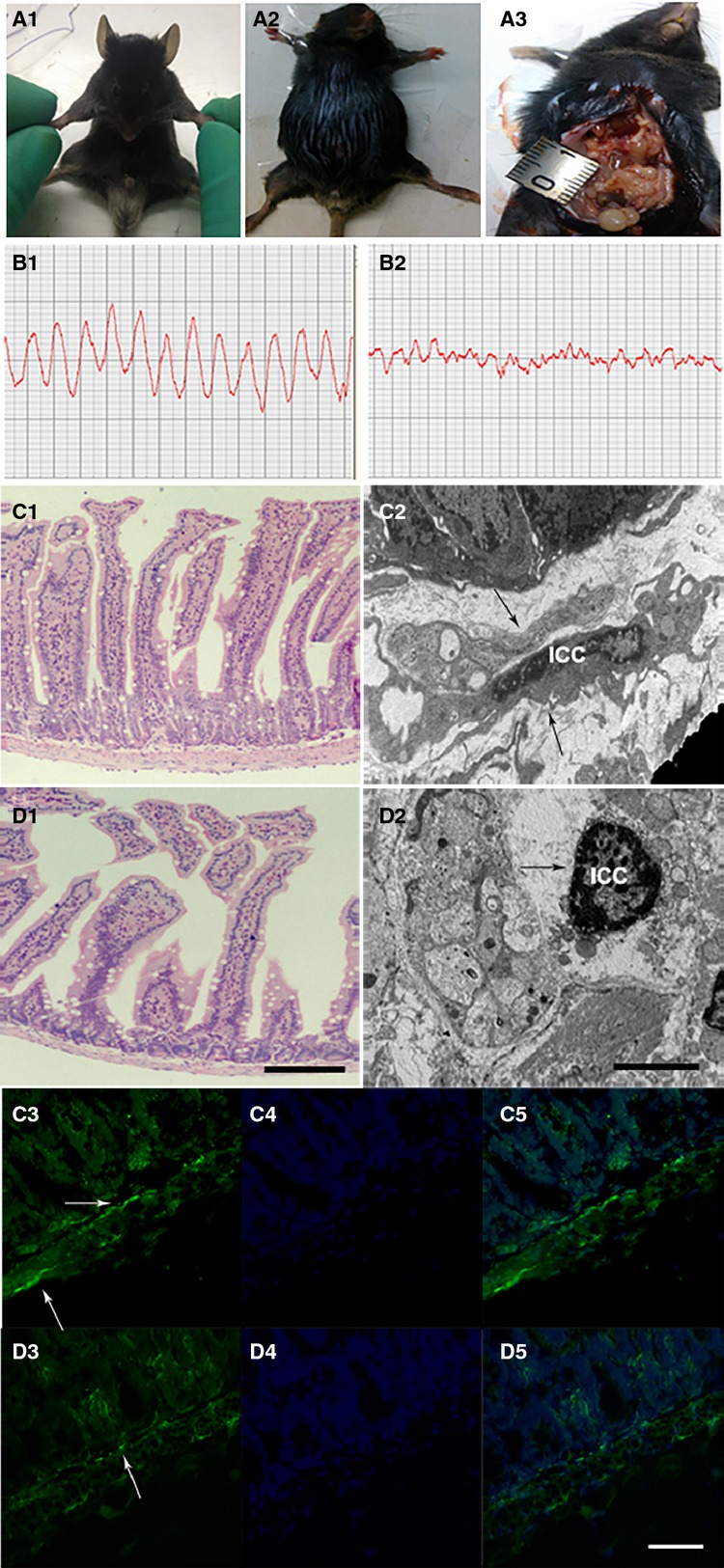
Figure 4.

The impact of malignant ascites on intestinal ICCs. (A1) Normal C57BL/6 mouse. (A2) Animal model creation of malignant ascites. (A3) The nodules in the abdominal cavity. Control mice exhibited regular waves and stable frequencies (B1), but in mice with malignant ascites, the amplitude of the intestine was clearly reduced and irregular (B2). On light microscopy, the integrity and columnar shape of the intestinal villi were maintained; the thickness of the muscularis propria was uniform (C1, LM ×200, bar: 250 μm), in the malignant ascites group, the intestinal villi were reduced and thinner, and the muscularis propria was significantly thinner than that in the control group (D1, LM ×200, bar: 250 μm). Ultrastructurally, ICCs displayed well-centred nuclei and numerous cytoplasmic processes that stretched and connected to nerve cells in control group (C2, black arrow, EM ×4000, bar: 12.5 μm), and in the malignant ascites group, the quantity and volume of ICCs were decreased, and many vacuoles were formed in the cytoplasm in the early stage of the model group. With the progression of disease, the nuclei clearly became condensed, the cell bodies became shrunken, and the cytoplasmic processes were decreased and had no connections with other cells (D2, black arrow, EM ×4000, bar: 12.5 μm). The immunofluorescence demonstrated that c-kit was partially expressed in the muscularis layer (C3, white arrow, IF ×400, bar: 125 μm), in the malignant ascites group, immunofluorescence showed few, spotty and scattered c-kit expression in the muscularis layer (D3, white arrow, IF ×400, bar: 125 μm). (C4 and D4) Nuclei are stained blue with DAPI. (C5 and D5) merged images.
