Figure 4.

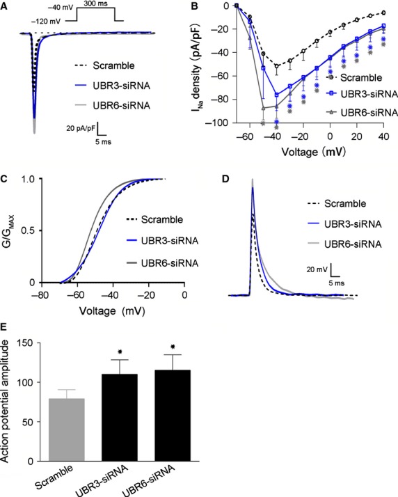
Effects of UBR3/6 knockdown on Nav1.5 channel currents and action potentials. (A) Representative tracings of Nav1.5 currents from rat cardiomyocytes. The amplitude of the UBR3/6 knockdown cells was increased. (B) Current–voltage (I–V) relationship of transient INa from rat cardiomyocytes (n > 10 per group *P < 0.01). The current traces were recorded at Vm in the range of −70 to +40 mV from a holding potential of −120 mV. The INa density of the UBR3/6 knockdown cells was elevated compared to the NC. (C) The activation curve for Nav1.5 channels from NC (negative control) and UBR3/6 knockdown rat cardiomyocytes (n > 10 per group). There were no significant differences between them. (D) Representative AP (action potential) recordings from NC (negative control) and UBR3/6 knockdown rat cardiomyocytes. (E) Statistical analysis of the amplitude of the APA (action potential amplitude) (n > 10 per group, *P < 0.01). The amplitude was increased in UBR3/6 knockdown cells.
