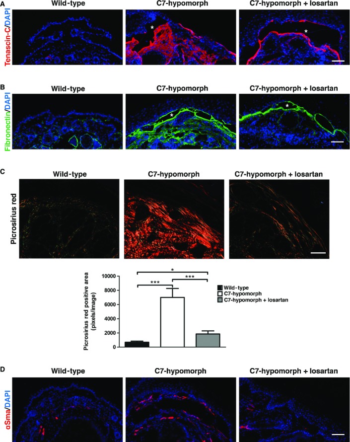
Figure 4. Reduced fibrotic remodeling in losartan-treated C7-deficient forepaws.
- A, B Tenascin-C (red) (A), fibronectin (green) (B). The nuclei were visualized with DAPI (blue). Images acquired with a 20× objective, scale bar = 100 μm. Note that losartan did not completely abolish the staining, but effectively limited fibrosis to the site of initial tissue damage, that is, adjacent to the dermal–epidermal blistering (denoted by white asterisks) in C7-hypomorphic skin.
- C Picrosirius red staining and visualization of the collagen fibers under cross polarizing light. Under this light, thin fibers appear green and thick rigid collagen bundles orange-red. The staining revealed significantly reduced collagen fiber size in losartan-treated skin, indicating softer tissue similar to wild-type skin. Below, the bar graph shows quantification of picrosirius red-positive areas, n ≥ 19 areas quantified, values represent mean ± S.E.M. Unpaired t-test with Welch’s correction, ***P-value wild-type vs. C7-hypomorph < 0.0001; ***P-value C7-hypomorph + losartan vs. C7-hypomorph = 0.0004; *P-value wild-type vs. C7-hypomorph + losartan = 0.0189. Images acquired with a 20× objective, scale bar = 50 μm.
- D Immunofluorescence staining of forepaws as above with an antibody to αSma (red). αSma is present both around blood vessels and in myofibroblasts. Note the increase of αSma+ myofibroblasts in C7-hypomorphic paws and reduced number of αSma+ cells in losartan-treated C7-hypomorphic forepaws. Images acquired with a 20× objective, scale bar = 100 μm.

