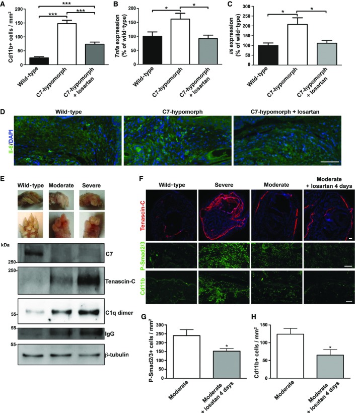
Figure 7. Tgf-β inhibition through losartan effectively relieves inflammation in RDEB.
- A Quantification of Cd11b-positive cells shows that losartan treatment significantly reduced the number of these cells in C7-hypomorphic paws. Values represent mean Cd11b-positive cells per 1 mm2 ± S.E.M. Unpaired t-test with Welch’s correction was used, ***P-value wild-type vs. C7-hypomorph < 0.0001; ***P-value C7-hypomorph + losartan vs. C7-hypomorph = 0.0008; ***P-value wild-type vs. C7-hypomorph + losartan < 0.0001 (n = 5).
- B, C qPCR analysis of Tnfa and Il6 mRNA expression in forepaws, normalized to the housekeeping gene Gapdh. Treatment with losartan significantly lowered the elevated expression of both genes in C7-hypomorphic paws. Values are expressed as percentage of expression in age-matched wild-type forepaws and represent mean ± S.E.M. Unpaired t-test with Welch’s correction used for data analysis. (B) *P-value wild-type vs. C7-hypomorph = 0.033; *P-value C7-hypomorph +losartan vs. C7-hypomorph = 0.015; P-value wild-type vs. C7-hypomorph + losartan = 0.57, n = 5. (C) *P-value wild-type vs. C7-hypomorph = 0.69; *P-value C7-hypomorph + losartan vs. C7-hypomorph = 0.029; P-value wild-type vs. C7-hypomorph + losartan = 0.57, n = 5.
- D Immunofluorescence staining of age-matched wild-type, untreated and losartan-treated C7-hypomorphic forepaws with an antibody to Il-6. The number of Il-6-positive bright cells (green) is clearly increased in untreated C7-deficient skin, and nearly normalized after 7-week losartan treatment. Nuclei visualized with DAPI. Images acquired with a 20× objective, scale bar = 100 μm.
- E Correlation of tissue inflammation with disease progression in RDEB. Photographs of wild-type and moderately and severely affected C7-hypomorphic forepaws. These paws were processed for Western blotting shown below. The blots were probed with antibodies detecting C7, tenascin-C, C1q, and IgG. β-tubulin was used as a loading control. Shown for C1q is a dimeric form (Wing et al, 1993). Note the difference between moderately and severely affected paws. The C7 expression does not differ, but the severely affected paw with more extensive fibrosis and tenascin-C expression indicating remodeling also displays more tissue inflammation, as shown by increased C1q and IgG levels.
- F Short-term losartan treatment rapidly alleviated inflammation through reduction of Tgf-β signaling. Sections of forepaws as in (E) plus sections of forepaws of C7-hypomorphic mice with moderately affected paws treated with losartan for 4 days were stained for tenascin-C, P-Smad2/3, and Cd11b. There is a clear correlation between the extent of fibrosis, as revealed by tenascin-C staining; Tgf-β signaling, as detected by P-Smad2/3; and inflammation, as indicated by Cd11b+ cells in the C7-hypomorphic forepaws. The 4-day losartan treatment efficiently reduced Tgf-β signaling (P-Smad2/3) and inflammation (Cd11b+) in moderately affected paws, as compared to untreated C7-hypomorphic paws with similar degree of fibrosis. Collectively, the data show that TGF-β-mediated inflammation is a driver of disease progression in RDEB and a major losartan target. Scale bars = 100 μm.
- G, H Quantification of stainings of moderately affected C7-hypomorphic forepaws with or without a 4-day losartan treatment as in (F). Positively stained cells were quantified after background had been subtracted by applying equal threshold. The values were expressed as positive cells per mm2. Values represent mean ± S.E.M. Data were analyzed with unpaired t-test with Welch’s correction. (G) P-Smad2/3 staining, *P = 0.033. (H) Cd11b+ cells, *P = 0.027. n = 3 different mice per group.
Source data are available online for this figure.

