
Fig 4.
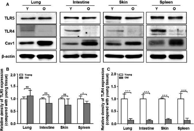
Expression levels of TLRs in various tissues from young and old mice. Tissues were isolated from young and aged mice. (A) The protein expression in the various tissues from young and aged mice was analyzed by Western blotting with anti-TLR5, anti-TLR4, anti-Cav1, and β-actin antibodies. The quantitative results of (B) TLR5 and (C) TLR4 expression in the various tissues are represented by the graphs. The relative density of proteins was normalized with β-actin. The data were based on the three independent experiments (n = 4 mice per each group). Differences were analyzed by Mann–Whitney U-test. *P < 0.05; ***P < 0.001; and ns, not significant compared with the tissues from the young mice.