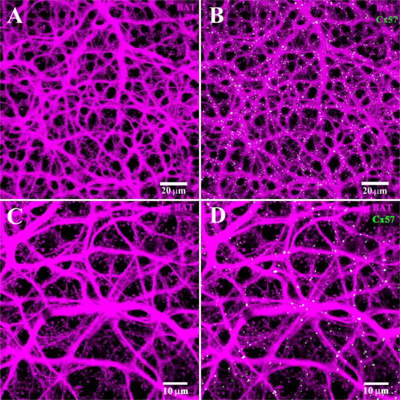
Figure 12.

B-type axon terminal plexus. A: The B-type AT plexus was filled with Neurobiotin (magenta). There are numerous fine endings which contact individual rod spherules and a complete absence of cell bodies. This indicates that the B-type AT plexus is labeled exclusively. B: The Cx57 plaques (green) are found colocalized with the B-type AT plexus. Note the Cx57 plaques appear almost exclusively white and there are almost no green, single-labeled plaques. C: High-magnification view of the AT plexus. The absence of B-type somas and the presence of numerous typical AT endings to match the distribution of rod spherules indicate that only the AT processes were labeled. D: Same frame, nearly all the Cx57 plaques are colocalized with the AT plexus.
