
Figure 13.
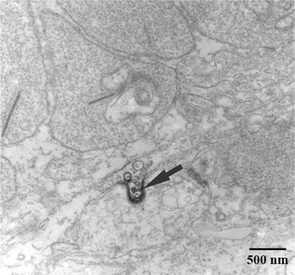
Electron microscopy for Cx57. The figure shows a region of the outer plexiform layer (OPL). The profiles at the top of the frame are rod spherules, as shown by the prominent synaptic ribbons and numerous synaptic vesicles. Cx57 immunoreactivity was found between two profiles. This putative Cx57 gap junction (arrow) has an invaginating profile. As previously noted for the confocal images, there was no evidence of Cx57 labeling associated with rod spherules.