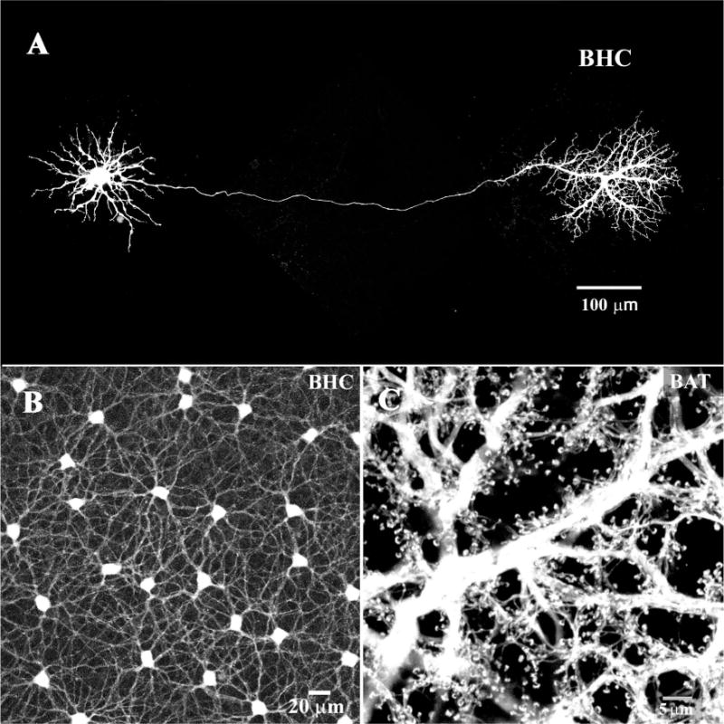
Figure 5.

B-type horizontal cells (HCs). A: A single B-type HC was dye injected with Neurobiotin in the presence of meclofenamic acid (MFA, 200 μM), to block gap junctions (Pan and Massey, 2007). Under these conditions, a single axon-bearing HC was obtained. The cell body, with somatic dendrites is on the left, connected by a very fine axon to the axon terminal, on the right. B: In the absence of MFA, when a B-type HC was dye injected the mosaic of dye-coupled somas was obtained connected by a plexus of somatic dendrites. C: In contrast, when an axon terminal was dye-injected a field of overlapping axon terminals was filled. Note the absence of cell bodies and the characteristic fine endings which contact individual rod spherules.
