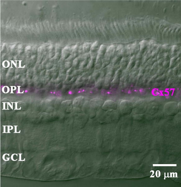
Figure 6.

Cx57 labeling of a cross-section of rabbit retina. In a DIC image, the layers of the rabbit retina were clearly visible. Staining with the mouse Cx57 antibody labeled bright clusters confined to the OPL. The rest of the retina was unlabeled.
