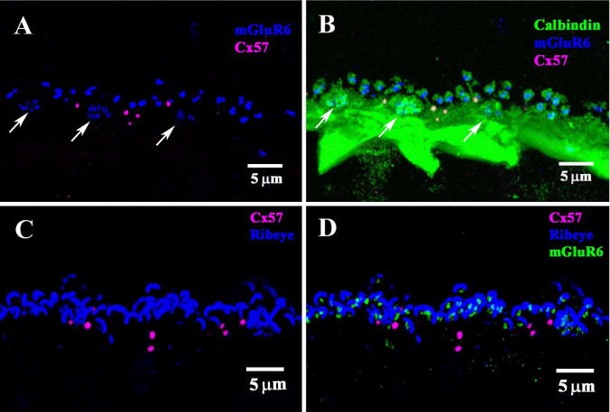
Figure 7.

Comparison of Cx57 labeling with other neuronal markers. A: Double label of vertical section of rabbit retina showed punctate labeling of Cx57 (magenta) in the OPL. Staining for mGluR6 (blue) produced clusters of fine ON bipolar terminals (arrows), which indicate the location of cone pedicles, and larger pairs of rod bipolar terminals, which show the location of rod spherules. The Cx57 plaques were below the level of the rod terminals, but occasionally above the level of the cone pedicles, as previously reported by Puller et al. (2009). This suggests an association with the ATs which also run at this level. B: Triple label image, with horizontal cells (HCs) stained with an antibody against calbindin (green). A very dense band was stained with a prominent cluster of HC terminals which converge at the site of each cone pedicle (arrows). Very fine HC processes left the plexus to contact each rod spherule, surrounding bright mGluR6 doublets. The Cx57 plaques lie at the top of the HC band mostly below the photoreceptor terminal. There was no apparent relationship between the Cx57 plaques and either cone pedicles or rod spherules. C: Double label image showing Cx57 plaques (magenta) and synaptic ribbons in the OPL stained for RIBEYE (blue). Many large curved rod synaptic ribbons were stained. D: Triple label image with mGluR6 receptors (green) nestled within each synaptic ribbon. As above, there was no apparent relationship between Cx57 and photoreceptor terminals. The Cx57 plaques were mostly below the level of photoreceptor terminals.
