
Figure 2.
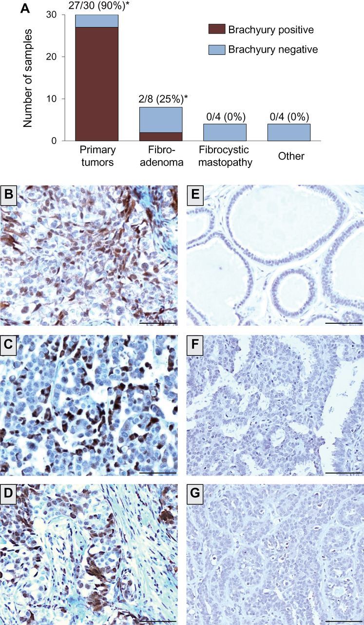
Brachyury protein expression in primary infiltrating ductal carcinomas vs benign breast tissues. A) Expression of brachyury protein was analyzed by immunohistochemistry with a brachyury-specific murine monoclonal antibody (Abcam) in 30 cases of primary infiltrating ductal carcinomas, eight cases of fibroadenoma, four cases of fibrocystic mastopathy, and one case of each of the following: usual ductal hyperplasia, sclerosing adenosis, intraductal papilloma, and normal terminal-ducto-lobular unity (TDLU), indicated in the graph as “other.” Shown is the number (and percentage) of brachyury-positive tissues per total of cases analyzed for each tissue type. (*) Two cases of adenocarcinoma and two cases of fibroadenoma scored as focal, defined as ≤5% of cells positive for brachyury. B–D) Transmitted light photomicrographs of representative primary infiltrating ductal carcinoma tissue sections stained for brachyury protein (B: patient 21; C: patient 24; D: patient 27 from Table 1). Also shown are representative examples of benign breast tissues negative for brachyury expression, corresponding to mastopathy (ductal ectasia) (E), usual ductal hyperplasia (F), and mastopathy (ductal epitheliosis) (G). The brown signal corresponds to brachyury. Magnification ×20. Scale bars = 100 μm.