
Figure 3.
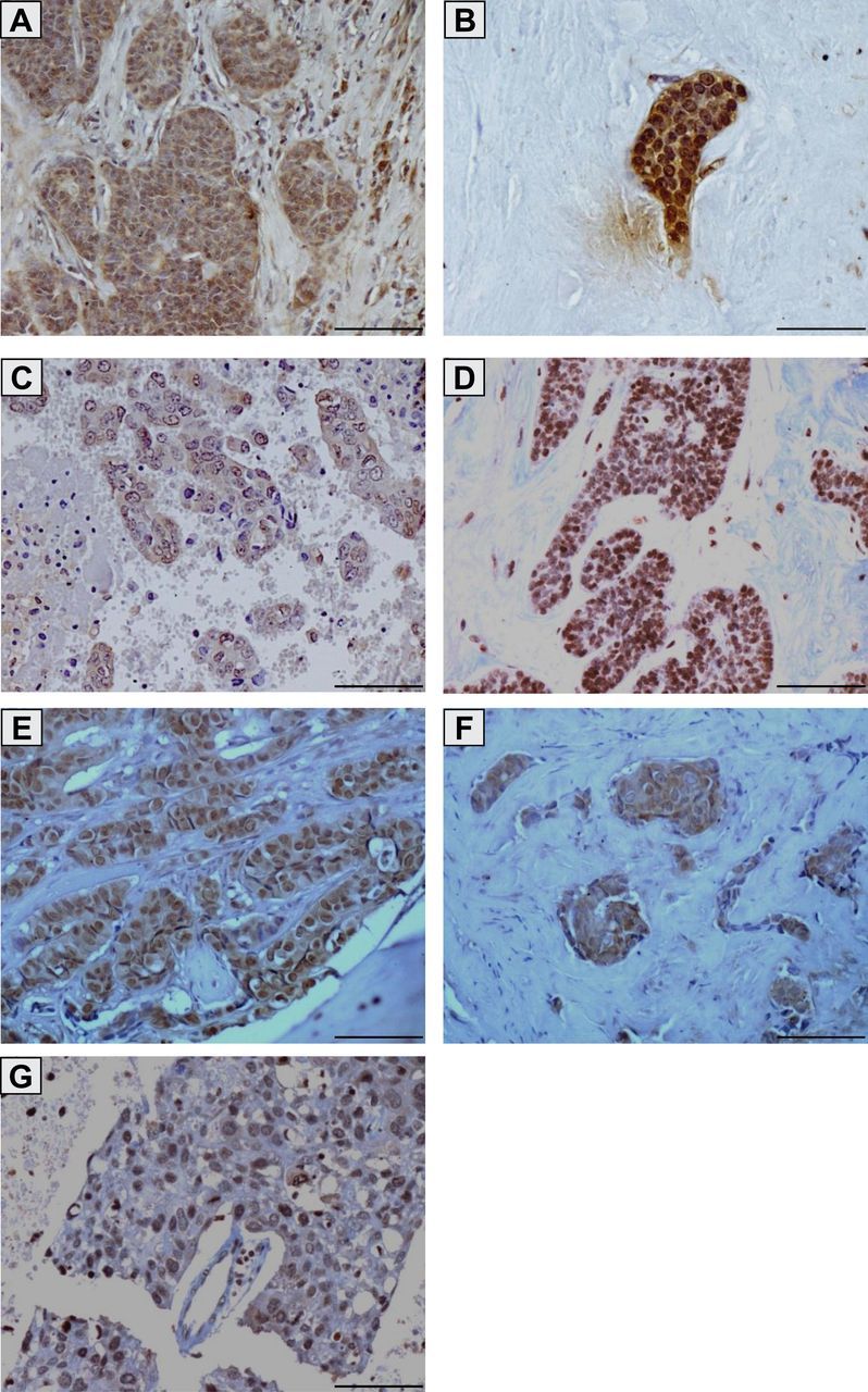
Brachyury protein expression in primary infiltrating ductal carcinomas and breast cancer metastases. Transmitted light photomicrographs of tissue sections stained for brachyury protein expression in a primary infiltrating ductal carcinoma (A) and a corresponding lymph node metastasis (B) from the same patient (patient 6 in Tables 1 and 2); a primary infiltrating ductal carcinoma (C) and a corresponding lymph node metastasis (D) from the same patient (patient 9 in Tables 1 and 2); bone metastatic lesions from two different breast cancer patients (E and F) (patients 32 and 33 in Table 2); and a brain metastatic lesion (G) (patient 34 in Table 2). The brown signal corresponds to brachyury. Magnification ×20. Scale bars = 100 μm.