
Figure 6.
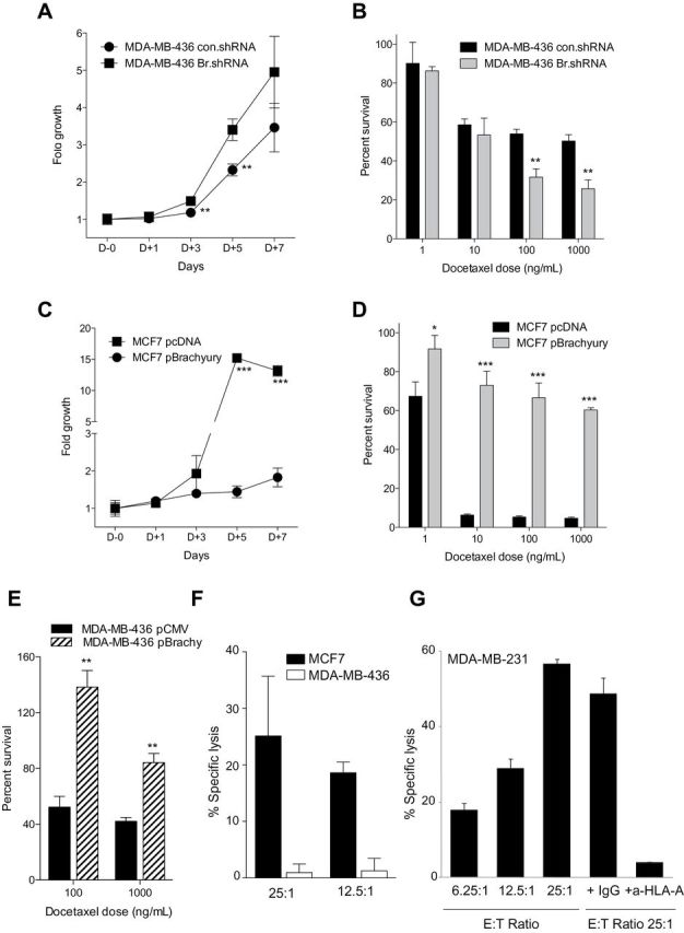
Brachyury-mediated tumor resistance and T-cell mediated cytotoxicity. MDA-MB-436 cells were transfected with a control nontargeting shRNA (con.shRNA) and a brachyury-specific shRNA (Br.shRNA). Tumor cells were evaluated for growth kinetics by the 3-(4,5-dimethylthiazolyl-2)-2,5-diphenyltetrazolium bromide (MTT) assay over a period of 7 days in culture (A) or 5-day survival in response to indicated doses of docetaxel (ng/mL) (B). C) MCF7 cells were transfected with an empty vector (pcDNA) or a Brachyury-encoding vector (pBrachyury) and evaluated by the MTT assay for growth kinetics over a period of 7 days in culture. D) The MCF7 tumor cell pair was treated with indicated doses of docetaxel (ng/mL) and assayed (day 5) for survival in comparison with untreated cells. E) Survival of MDA-MB-436 cells transfected with a control pCMV vs pBrachyury vector in response to indicated doses of docetaxel. F) Brachyury-specific cytotoxic CD8+ T cells expanded in vitro from the blood of a prostate cancer patient by using a human leukocyte antigen A2 (HLA-A2) binding 9-mer peptide of brachyury were able to lyse MCF7 cells that are brachyury positive and HLA-A2 positive, but not the MDA-MB-436 cells that are brachyury positive but HLA-A2 negative. G) An additional breast carcinoma cell line, MDA-MB-231, which is brachyury positive and HLA-A2 positive was also used; brachyury-specific T cells were able to efficiently lyse MDA-MB-231 cells at different effector-to-target cell (E:T) ratios. Tumor lysis was almost completely abrogated by the addition of an anti-HLA-A antibody but not a control immunoglobulin G (IgG). In all panels, bars represent the mean ± standard deviation of at least three identical replicates. Results in panels C, D, E and G are representative of at least two independent experiments. *P < .05, **P ≤ .005, ***P < .001 by Student t test analysis.