
Fig. 29.
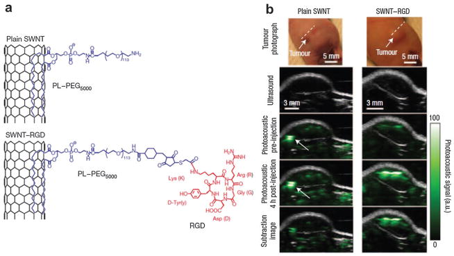
(a) Schematic illustrating SWNTs conjugated with RGD peptides for targeted PAT of mouse tumor.169 (b) B-scan US and PA images of U87MG tumor acquired along a white dotted line aided by SWNTs.169 The US images (gray) show the skin and tumor boundaries, while PAT images (green) show optical absorption (SWNT–RGD) in the tumor. Differential images were obtained by subtraction of the pre-injection image from the 4 h post-injection image.