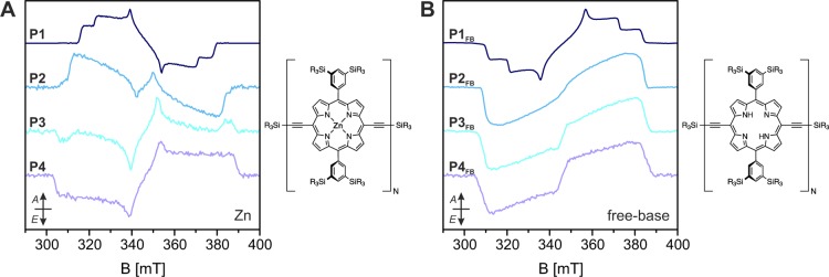
Figure 3.

Experimental X-band transient EPR spectra of the zinc (A) and free-base (B) linear porphyrin oligomers P1–P4 in MeTHF:pyridine 10:1 recorded as average up to 2 μs after the laser pulse at 20 K. The spectra were recorded after excitation at wavelengths corresponding to the planar conformations (645, 750, 800, and 830 nm for the zinc porphyrins and 680, 740, 780, and 810 nm for the free-base porphyrins, see UV–vis data in the Supporting Information). At shorter wavelengths, the contribution of different conformations affects the spin polarization of the EPR spectrum.
