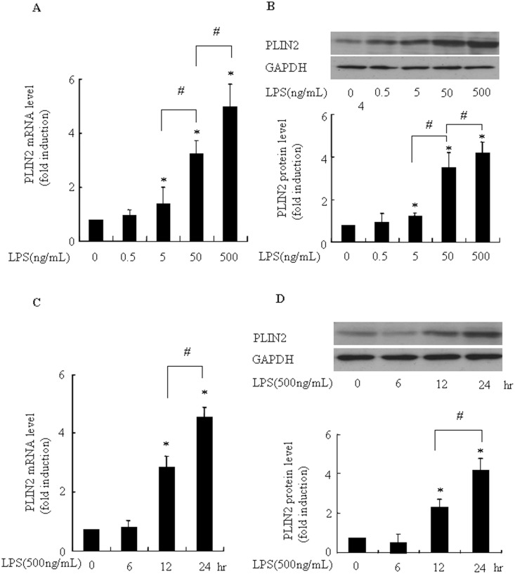
Fig 6. LPS increased the levels of PLIN2 mRNA and protein in a dose and time dependent manner in BV2 microglia.
(A and B): LPS stimulated PLIN2 mRNA and protein expression in a dose-dependent manner. Cells were incubated with vehicle or indicated concentrations of LPS for 24 h. (C and D): LPS enhanced PLIN2 mRNA and protein expression in a time-dependent manner. Cells were incubated with vehicle or LPS (500ng/mL) for 6, 12 or 24h. Ribosomal RNAs and GAPDH were used as the total RNA or protein loading control, respectively. The PLIN2 mRNA level in the control (no stimuli) was arbitrarily designated as 1 for comparison. Levels of PLIN2 protein were quantified by the NIH Image processing and analysis program. *P< 0.05 compared with LPS alone. #P< 0.05. Experiments were repeated 3 times and representative results are shown.

