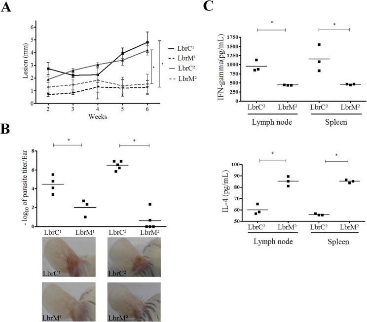
Fig 3. LbrC and LbrM isolates exhibit distinct virulence in BALB/c mice.
(A) The panel represents the lesion development in hamsters (Mesocricetus auratus) after infection with the isolates. Ear thickness was measured with a Mitutuyo digital caliper for six weeks. Each point represents the mean lesion size (±SEM) of five animals per group. (B) The graph represents the parasite load in the ear of BALB/c mice after a four-week infection with the isolates by limiting dilution assay. Three or five mice were used per group. The lower panel shows representative images of ear lesions after 4 weeks of infection. (C) Quantification of IFN-γ and IL-4 cytokines released by lymph node and spleen cells after one month of infection with the LbrC2 and LbrM2 isolates following stimulation with SLA for 72 h. Three mice were included in each group. *p<0.05 (two-tailed t-test).

