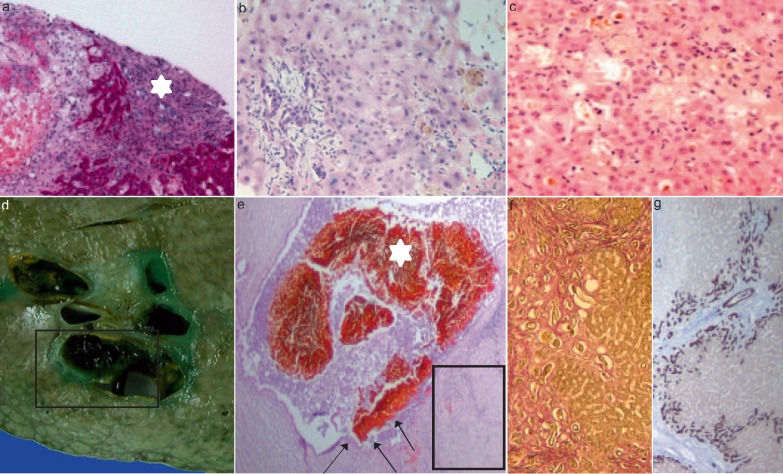
Fig. 2.
Liver biopsy samples showing morphological features of SC-CIP (portal and parenchymal changes); a-c portal tract showing marked enlargement due to edema, mild mixed inflammatory infiltrate, and mild fibrosis. Adjacent, a periportal bile duct infarct (a, asterisk; periodic acid-schiff stain, 100×) can be seen. Degenerative changes of the bile duct epithelium which included loss of cellular polarity, cellular dropout, and irregularities of the basal membrane (b, hematoxylin and eosin stain, 200×). Hepatocellular and canalicular cholestasis (c, hematoxylin and eosin stain, 200×). Explanted liver: Gross appearance (d) and histological section (e, f) showing severe damage to the large bile duct with cholangiectasis, intraductal bile sludge as well as biliary casts (asterisks; square in d), and segmental ulceration (arrows) of the bile duct epithelium (e, hematoxylin and eosin stain, 10×). Secondary biliary cirrhosis (f, g): progressive periportal and septal fibrosis with bridges linking adjacent portal tracts (f, EVG, 100×; square in e). Marginal bile duct proliferation and ductular metaplasia of periportal hepatocytes with strong expression of cytokeratin 7 (g, 50×; square in e).

