Figure 1.
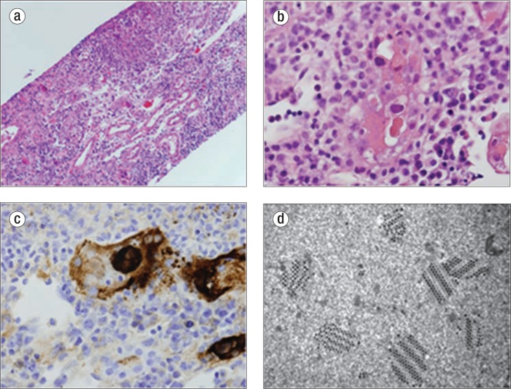
Renal biopsy. (a) Severe adenovirus interstitial nephritis in a kidney allograft consisting of tubular necrosis and brisk interstitial inflammation, hematoxylin and eosin (H&E) ×200. (b) Smudgy basophilic nuclear inclusion within a necrotic tubule, which is indicative of a viral inclusion, H&E ×400. (c) An immunoperoxidase stain with an antibody to adenovirus revealing positive staining in both the nucleus and cytoplasm of an infected cell, ×400. (d) Electron microscopy crystalline array of virus particles within a tubular epithelial cell, ×42,000.

