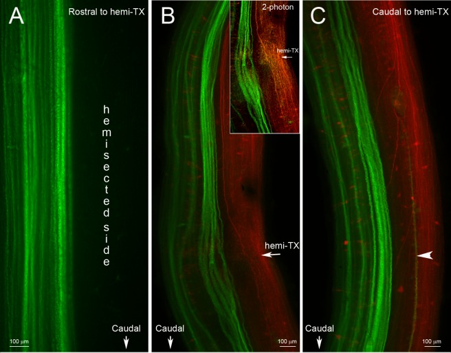
Fig 1. No collateral sprouting from spared spinal cord axons at 4 or 5 weeks after a hemi-TX.
A, unilateral labeling of previously uninjured axons in a wholemounted spinal cord rostral to a right-sided hemi-TX. Four weeks after the hemi-TX, a green dye (DAF 488) was applied to a complete TX 1 cm caudal to the hemi-TX. Axons severed by the hemi-TX were not labeled, while the originally non-injured axons were labeled in green. No decussating sprouts are seen. B and C, wholemounted spinal cord of an animal rostral (B) and caudal (C) to the hemi-TX site at the level of the 5th gill (arrow in B). DTMR (red) was applied to the hemi-TX, labeling the severed axons and their cell bodies. Five weeks later, a complete TX was made 1 cm caudal to the hemi-TX and DAF 488 (green) was applied, labeling the previously spared axons. The inset in B is a 2-photon photomicrograph of the hemi-TX region, showing axons regenerating across the hemi-TX. B and C show no evidence of collateral sprouting by the green-labeled (previously spared) axons. A single longitudinal green fiber in C (arrowhead) is the decussated rostrally-projecting axon of a giant interneuron whose perikaryon is located caudal to the depicted level.

