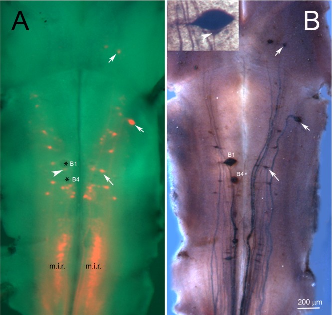
Fig 5. Surviving neurons continue to express NF180 protein after axotomy.

In an animal 10 months post-TX at the level of the 5th gill, DTMR was applied to a second complete TX at the level of the 3rd gill (approximately 5 mm below the obex of brain and 2 mm rostral to the original TX site), in order to label neurons projecting to the spinal cord, regardless of whether their axons had regenerated. After 5 days to allow for retrograde labeling, the brain was removed, pinned flat, and photographed live before fixation. A, fluorescently-labeled RNs superimposed on a brightfield image of the living brain. B, After fixation, the same brain was stained for NF180. DTMR labeling correlated closely with NF180 immunostaining, but there were exceptions: 1) Two swollen neurons (* in A; probably the left B1 and B4) were labeled by anti-NF180 but were not backfilled by DTMR, indicating that they had survived but their axons had retracted long distances and had not regenerated back to the level of the 3rd gill. 2) Some neurons were labeled by DTMR but not by NF180. Prominent among these were neurons in the medial inferior reticulospinal group (m.i.r.), reflecting that not all spinal-projecting neurons express NF180 in animals of this age [17]. The faint stain just caudal to B1 (white arrowhead in A) is a small neuron, not collapsed B1 cytoplasm, as shown in the inset of B. The white arrows in A and B point to neurons that are double-labeled because they have axons that regenerated and also express NF180.
