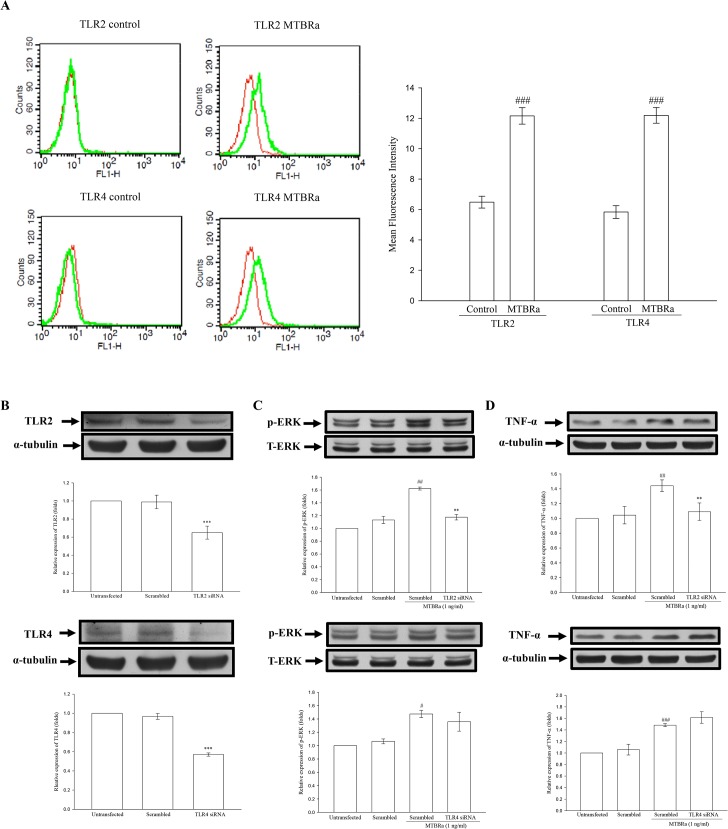
Fig 5. TLR/ERK Signaling Activating TNF-α Expression in MTBRa-Stimulated Human PMCs.
(A), Flow cytometric analyses of expression of TLR2 or TLR4 (green) on cell surface of MeT-5A cells treated with MTBRa (1 ng/ml) for 3 h or left untreated (control). Isotype control IgG (red). Quantitative analysis of TLR2 or TLR4 expression was expressed as percentage of the total cells. A representative of three experiments is depicted. ###p<0.001 compared with the control group. MeT-5A cells were transfected with scrambled siRNA, TLR2 siRNA (25 nM) or TLR4 siRNA (25 nM). After 30 min, 3 and 6 h of stimulation with MTBRa, the cellular extracts were prepared and the protein amounts of (B), TLR2 and TLR4, (C) ERK, and (D), TNF-α were determined by Western blotting. The data represent four independent experiments. ##p <0.01, ###p<0.001 compared with the control (scrambled RNA) group; **p<0.01, ***p<0.001 compared with scrambled RNA-transfected MTBRa-treated group. PMC, pleural mesothelial cell; TLR, toll-like receptor; MTBRa, heat-killed M. tuberculosis H37Ra; ERK, extracellular-signal-regulated kinase; TNF, tumor necrosis factor.

