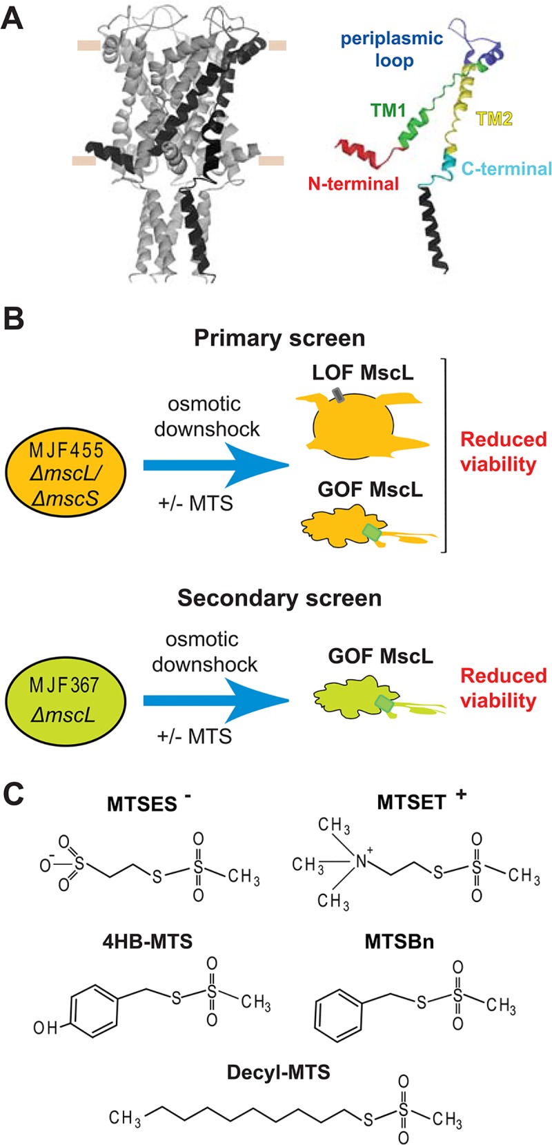
Fig 1. In vivo screens to determine MscL activity changes after post-translational modifications.

(A) The structure of E. coli MscL in its closed state [16], generously provided by Ben Corry, is shown in a side view, with a subunit highlighted for clarity. The approximate location of the lipid membrane headgroups is marked by horizontal tan lines. The domains, in which the study has been divided, are highlighted in an isolated subunit using different colors. (B) A schematic description of the in vivo screens used to study the activity of the MscL cysteine substituted channels before and after post-translational modification with different MTS reagents. In a primary screen, the osmotically fragile strain MJF 455 is osmotically shocked with or without the MTS reagents present during the osmotic down-shock. Channels with reduced sensitivity (Loss of function LOF) or increased sensitive to tension (gain of function GOF), lead to cells with reduce viability in the primary in vivo screens. A secondary screen is done to distinguish between these two phenotypes. The secondary screen consists in MJF367 strain (not osmotically fragile) osmotically shocked with the MTS reagents present in during the osmotic down-shock. In the secondary screens a reduced viability indicates more sensitive channel or GOF. (C) The structure of the sulfhydryl reagents 2-sulfonatoethyl methanethiosulfonate sodium salt (MTSES-), ethyl methanethiosulfonate Bromide (MTSET+), 4-hydroxybenzyl methanethiosulfonate (4-HB-MTS), benzyl methanethiosulfonate (MTSBn), and decyl methanethiosulfonate (decyl-MTS), 2-(Trimethylammonium) are shown.
