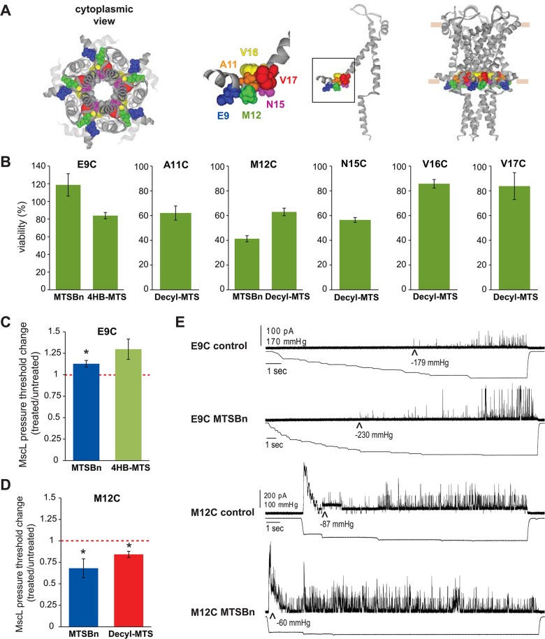
Fig 3. Functional changes by substitutions in the N-terminal domain of MscL, determined by in vivo and patch clamp experiments.
(A) The location of the residues showing changes in viability upon post-translational modifications is highlighted in the closed structure of E. coli MscL. From right to left: a pentameric MscL is shown in a side view followed by a single subunit and a close up of the region and a cytoplasmic view of the pentameric channel. (B) Viability of MJF 367 (mscL -) shocked in the presence of the indicated MTS reagent is shown for each individual MscL mutant. (C) The changes in the pressure threshold required to gate E9C MscL caused by treatment with MTS reagents is graphed as the ratio between before and after modification of the same patch. The red line indicated no change. (D) The changes in the pressure threshold required to gate M12C MscL caused by MTS reagents is graphed as the ratio between before and after modification of the same patch. The red line indicates no change. (E) Representative traces of E9C MscL and M12C before (control) and after treatment with MTSBn. The upper traces correspond to the current and the lower traces the negative pressure applied to the patch.

