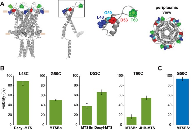
Fig 7. Functional changes by substitutions in the periplasmic loop of MscL, determined by in vivo and patch clamp experiments.
(A) The location of the residues showing changes in viability upon post-translational modifications is highlighted in the closed structure of E. coli MscL. From right to left: a pentameric MscL is shown in cytoplasmic view, a periplasmic view, and a side view where the approximate location of the membrane is shown. A single subunit and a close up of the region with the labeled residues is on the left. (B) Viability of MJF 367 (mscL -) shock in the presence of the indicated MTS reagent is shown for each individual MscL mutant, for hydrophobic or (C) charged MTS reagents.

