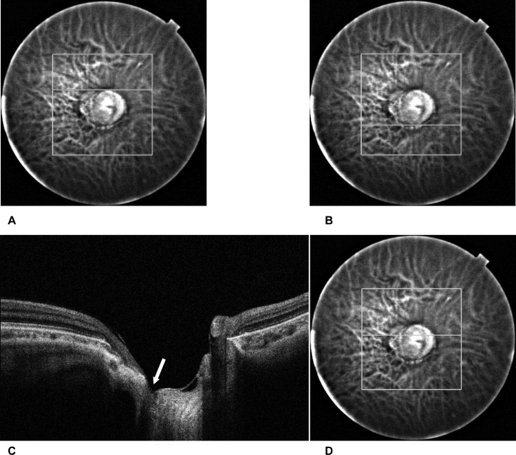
Fig 1. Identification of the defect location.
A) The superior disc edge identified on a fundus image obtained with the SS-OCT in reference to the color fundus photograph. In this case, the superior disc edge was located at the 90th scan of the sequential horizontal scans. B) The inferior disc edge was identified in a way similar to the superior edge. In this case, the inferior disc edge was located at the 180th scan. This means the center of the disc is located at the 135th scan. C) The location of the defect. A defect of the lamina cribrosa was identified in the OCT image (arrow). In this case the defect was located from 115th through 118th scan, which means the defect location is in the superior half of the disc. D) In the reference fundus image, a horizontal white line shows the location of the scan.

