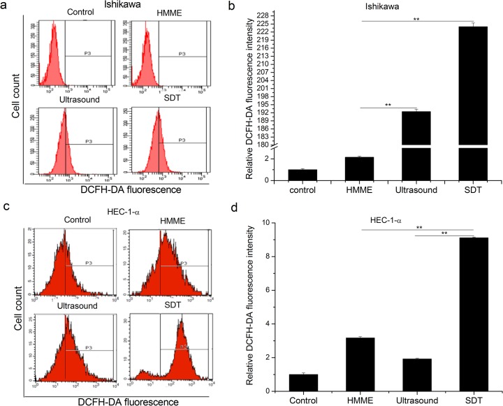
Fig 5. Effect of SDT on the ROS generation in endometrial cancer cells.
ROS levels are significantly enhanced in SDT groups when compared with the control, HMME, and ultrasound groups in Ishikawa (a and b) and HEC-1-a (c and d) cells. Representative FACS profiles were shown in a and c. Histograms present mean ± SD of three independent experiments (b and d, ** P < 0.01.)

