FIGURE 3:
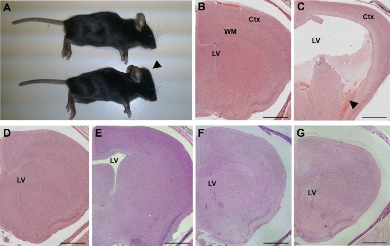
Hydrocephalus in Cfap54gt/gt mice. (A) WT (top) and Cfap54gt/gt (bottom) mice. The enlarged cranial vault (arrowhead) observed in mutant mice is suggestive of hydrocephalus. (B–G) Coronal histological sections of B6 WT (B), B6 Cfap54gt/gt (C), 129 WT (D), 129 Cfap54gt/gt (E), (B6×129)F1 WT (F), and (B6×129)F1 Cfap54gt/gt (G) brains through the lateral ventricles showing one hemisphere. There is dramatic dilatation of the lateral ventricles (LV) in the B6 mutant brain, as well as loss of the ciliated ependymal cells, damage to the white matter (WM) and cerebral cortex (Ctx), and hemorrhaging (arrowhead) (C). Only mild dilatation without substantial secondary tissue damage is observed in 129 (E) or (B6×129)F1 (G) Cfap54gt/gt mutant brains. Sections are stained with H&E. Scale bars: 1 mm.

