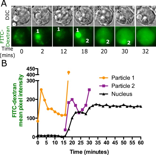
FIGURE 2:

FITC-dextran leakage occurs only from a silica-containing phagolysosome. A representative series of images (A) and quantification of the data (B) from a cell loaded with 4- kDa FITC-dextran for 2.5 h and then exposed to a small number of silica particles so that each particle could be tracked. Images were captured 2 min apart on a wide-field epifluorescence microscope. Each particle goes through the same sequence described in Figure 1. Particle 1 showed an initial increase in fluorescence and then a decrease as the FITC-dextran was quenched. At the time of leakage, the phagosome became transiently bright and then dimmer, whereas the nuclear fluorescence increased with a slight delay. Particle 2 showed the same pattern at a later time. Each particle seems to leak only once, but each time a phagosome shows this pattern, there is a correlated increase in nuclear fluorescence. The same quantal changes in nuclear and phagosomal fluorescence were observed in 33 particles in 20 cells.
