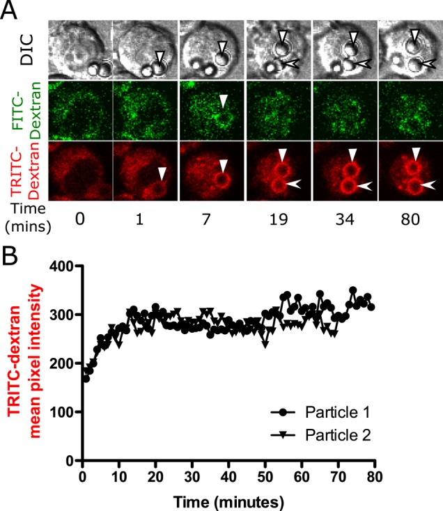
FIGURE 3:

Exposure to latex particle does not result in phagolysosomal leakage. Alveolar macrophages were loaded with 4-kDa FITC-dextran and TRITC-dextran (A, B) for 2.5 h, after which they were exposed to 20 μg/cm2 opsonized 3-μm latex particles. Volumetric image stacks were captured 2 μm apart every minute. (A) A sequence of representative images and (B) quantification of the images. Latex particles are not porous, so the phagosomal fluorescence appears as a ring instead of an entire volume of circle filled with fluorescence. Phagosomes containing latex particles show the same pattern of fluorescence changes as silica phagosomes up to the point of leakage. A brief increase in FITC-dextran fluorescence is observed around the particle at 7 min (marked by solid triangle) before phagosome acidification. (B) Particle 1 (solid triangle) and particle 2 (arrowhead) represent quantified data of particles in the TRITC-dextran channel. These trends are representative of 20 cells examined.
