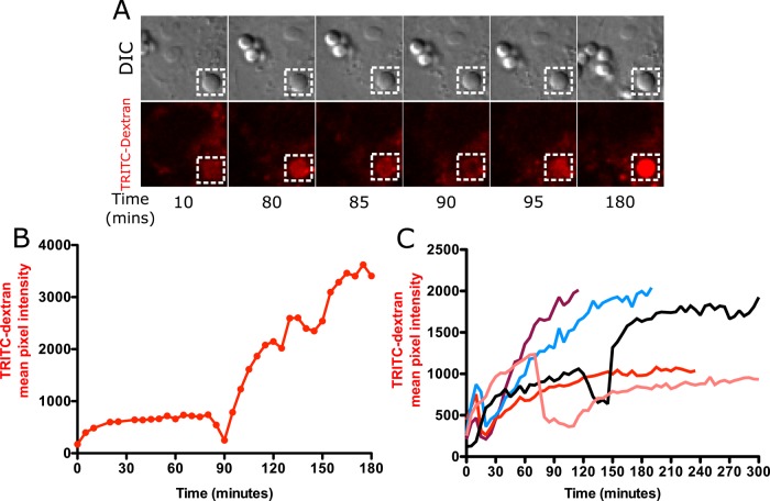
FIGURE 6:
Phagolysosomal leakage occurs in Cos7 cells exposed to silica particles. (A) Cos7 cells loaded with 4-kDa TRITC-dextran were exposed to 20 μg/cm2 nonopsonized silica particles and imaged at 5-min intervals. Note that Cos7 cells take a long time to initiate the process of phagocytosis after exposure to particles. Images were therefore acquired at 5-min intervals to prevent any inhibition of particle uptake due to excessive photoexposure. The phagolysosomal leakage data for Cos7 cells therefore lack a comparable temporal resolution to that of MH-S cells, where particle uptake is rapid and images were acquired 20 s apart. A gradual increase in TRITC-dextran fluorescence was observed upon particle phagocytosis (10 and 80 min), followed by a decrease in fluorescence starting at 85 min, with maximum loss of signal observed at 90 min. Vesicle refusion starts at 95 min, leading to a subsequent gradual increase in TRITC fluorescence. (B) Quantification of the phagosome in A. (C) Phagolysosomal leakage of individual phagosomes from different cells.

