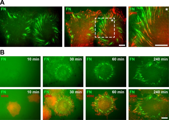
FIGURE 1:
Cell-induced FN fibrillogenesis. (A) TIRF microscopy images of REF52 cells incubated on a homogeneous coating of Alexa 488–labeled FN (FN-AF488, green) for 4 h and stained for vinculin (red) to visualize focal adhesions. A magnified view (rightmost image) corresponding to a boxed area of the merge image demonstrates the close association of FN fibrils and focal adhesions at the cell periphery. Scale bars, 10 μm. (B) Dynamics of cell-induced FN fibrillogenesis. REF52 cells were incubated on FN-AF488 (green) for 10, 30, 60, or 240 min, fixed, and stained for vinculin (red). Scale bar, 10 μm.

