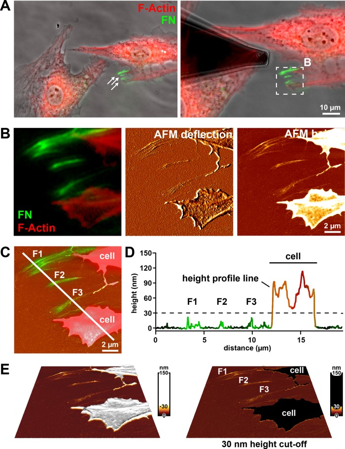
FIGURE 2:
Imaging FN fibrils using combined AFM and fluorescence microscopy (A) Left, phase contrast image with overlaid fluorescence signals for F-actin (red) and FN (green) of fixed Ref52 cells after 4 h of incubation on FN-AF488. The epifluorescence images were collected using an Achroplan 40×/0.80 W water-immersion lens. White arrows indicate assemblies of FN fibrils in the cell vicinity. Right, the same cell group with an AFM cantilever positioned near the fibrillar FN array. The white box indicates an area imaged by AFM and shown at higher magnification in B. (B) Merged fluorescence image (F-actin in red, FN in green) and corresponding AFM height and deflection mode images. (C) AFM height image overlaid with the FN fluorescence channel and the F-actin signal to indicate cellular protrusions. A profile line crosses several fibrillar FN arrays (F1, F2, F3) and a cellular protrusion. (D) Superimposition of height and fluorescence intensity profiles generated along the white line in C. FN fibrils (F1, F2, F3) can be distinguished from cellular protrusion or retraction structures (“cell”) using a 30-nm height cut-off (dashed line). (E) Three-dimensional reconstruction of the AFM height image shown in B, applying either a nonlinear height scale (left) or a 30-nm height cut-off (left), which eliminates all cellular structures (“cell”) from the image but retains all fibrillar FN structures (F1, F2, F3).

