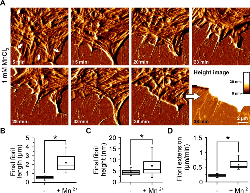
FIGURE 5:
Enhanced early fibrillogenesis in presence of Mn2+. Cells were incubated for 5 min on FN in the presence of Mn2+ (1 mM) and then continuously imaged by AFM. (A) FN fibrils are already visible after 5 min (white arrows) and show enhanced growth rates. Scale bar, 1.5 μm; full range of the AFM height scale, 20 nm. (B) Box-and-whisker plot of final fibril length in absence or presence of 1 mM Mn2+. (C) Box-and-whisker plot of final fibril height in absence or presence of 1 mM Mn2+. (D) Velocity of lamellipodium retraction in absence or presence of 1 mM Mn2+. Data (mean ± SD) from nine independent experiments. Statistically significant differences (p < 0.01) are denoted by an asterisk. The complete time-lapse series is presented in Supplemental Movie S2.

