FIGURE 6:
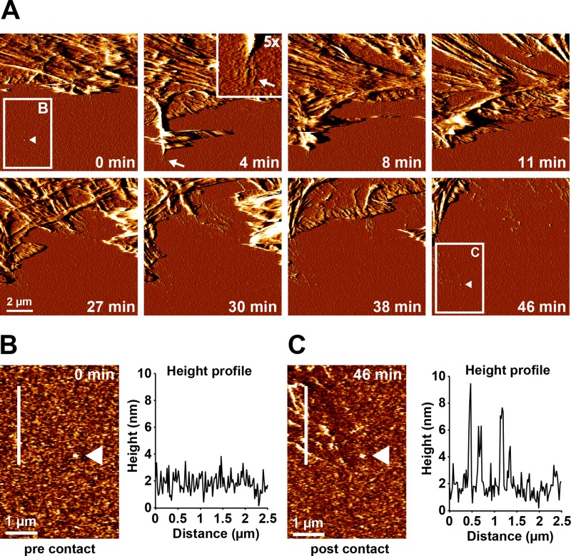
Fast FN rearrangement at retracting cell membranes. Cells were adhered to a homogeneous FN substrate in the presence of 1 mM Mn2+ for 10 min. Subsequently, a 10 × 10 μm2 area at the cell edge was continuously imaged by AFM in contact mode. (A) Time series of AFM deflection images showing part of a cell lamellipodium next to an uncontacted area on the FN surface. After 4 min, a transient cellular extension first forms and then retracts, inducing the formation of FN nanofibrils in the process (arrow). Inset, magnified view (5×) of the tip of the cellular extension and the associated FN nanofibrils. After several rounds of extension and retraction (8–30 min), the cell gradually retracts out of the imaging area, leaving behind a remodeled FN layer. Higher-resolution AFM height images of the region indicated by the white rectangle in A before cellular contact at time point zero (B) and after complete cell retraction 46 min later (C). A height profile along the white line demonstrates only small (<3 nm) variations in FN height before cellular contact (B), but large (≤10 nm) variations in FN height after retraction, consistent with the formation of FN nanofibrils. White triangles points to a small irregularity in the FN layer used as a positional marker in both images. The entire time-lapse series is presented in Supplemental Movie S3.

