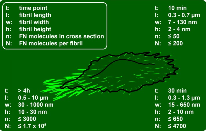
FIGURE 8:
Time line of early cell-induced FN fibrillogenesis. Fibrillar volumes were approximated from fibrillar height, width, and length values extracted from AFM height images. Width values were corrected for tip convolution (see Supplemental Figure S6). Number of FN molecules per fibril cross section and total molecules per fibril were estimated assuming maximal hexagonal packing of cylindrical FN dimers with a length of 160 nm, a diameter of 3 nm, and a 90-nm stagger between dimer building blocks (see Supplemental Figure S6).

