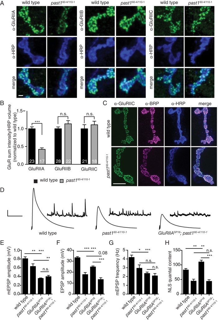
FIGURE 2:
Past1 mutants have reduced postsynaptic responses and defective homeostatic compensation. (A) Past1 mutants have reduced GluRIIA levels but normal GluRIIB and GluRIIC levels. Maximum intensity projections of 60× spinning-disk confocal stacks from a representative muscle 4. Scale bar, 2 μm. (B) Quantification of GluR levels from 3D volumes surrounding HRP staining. (C) Normal presynaptic (α-BRP) and postsynaptic (α-GluRIIC) apposition in past1 mutants. Maximum intensity projections of 100× spinning-disk confocal stacks from a representative muscle 4. Scale bar, 10 μm. (D) Representative traces from muscle recordings. The x-axis scale bar, 50 ms (EPSPs), 10,000 ms (mEPSPs); y-axis scale bar, 5 mV (EPSPs), 1 mV (mEPSPs). (E–H) Quantification of electrophysiological phenotypes. (E) mEPSP amplitude, (F) EPSP amplitude, (G) mEPSP frequency, and (H) quantal content, adjusted for nonlinear summation (NLS). Genotypes include white (wild type), n = 31; past160-4/110-1, n = 13; GluRIIASP16, n = 44; and GluRIIASP16; past160-4/110-1, n = 16.

