FIGURE 3:
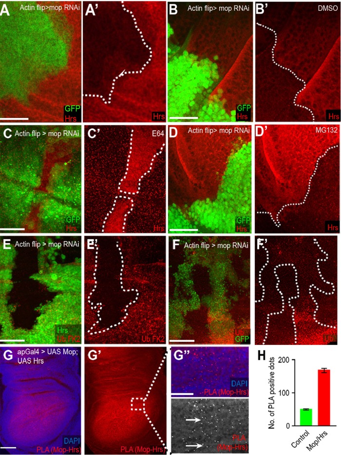
Mop protects Hrs from lysosomal degradation. (A, A′) Hrs is reduced in mopRNAi clones (marked with GFP). (B, B′) mopRNAi disks treated with DMSO. (C, C′) Disruption of lysosomal degradation by E64 treatment prevents Hrs degradation, whereas inhibiting proteasomal degradation by MG132 has no effect (D, D′). (E, E′) Knockdown of mop results in increased ubiquitinated proteins and monoubiquitins and polyubiquitins (F, F′). (G–G′′) PLA between Mop and Hrs shows a strong association between the two (G′′, arrows). (H) Quantification of PLA red dots in control and Mop-Hrs–overexpressing tissue shows an ∼3.5-fold increase in Mop–Hrs association in the affected tissue. mop was knocked down using VDRC line 104860. Scale bars, 20 μm.

