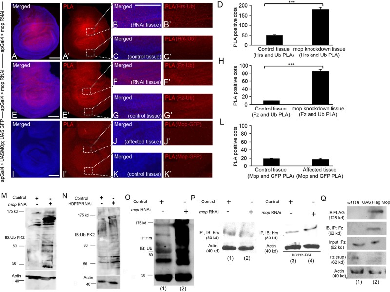
FIGURE 4:
Hrs and Fz show increased association with ubiquitin in the absence of Mop. In situ PLA in Drosophila wing disks expressing (A, A′) mopRNAi in dorsal half of wing pouch using ap-Gal4 to visualize association between Hrs and Ub. Boxed regions in A′ were zoomed in to show affected (B, B′) and control (C, C′) tissue. PLA red dots were more abundant in B and B′ than in C and C′. (D) Quantification of the dots suggests increased complex formation between Hrs and Ub upon mop knockdown (p < 0.003). (E, E′) In situ PLA of Fz and Ub in disks expressing mopRNAi. Boxed regions in E′ were zoomed in to show affected (F, F′) and control (G, G′) tissue. PLA red dots were more abundant in F and F′ than in G and G′. (H) Quantification of the dots indicates increased association between Fz and Ub upon mop knockdown (p < 0.001). (I, I′) In situ PLA of Mop and GFP. Boxed regions in I′ were zoomed in to show affected (J, J′) and control (K, K′) tissue. PLA red dots were not changed in J and J′ compared to K and K′. (L) Quantification of the dots shows no change in red dots in each case (p < 0.19). Scale bars, 20 μm. (M) Knockdown of mop in Drosophila wing disks causes increased levels of ubiquitinated proteins. mop was knocked down using VDRC line 104860. (N) Knockdown of HDPTP causes an increase in ubiquitinated protein levels in mouse L cells. (O) Protein lysates from Drosophila wing disks were assayed for levels of ubiquitin in control and mop-knockdown tissue using MS1096Gal4. Immunoprecipitation assays were performed with anti-Hrs antibodies, and extracts were visualized by Western blotting by using anti-Ub FK2 antibodies. (P) Immunoblotting of Hrs after Hrs immunoprecipitation in control and mop-knockdown tissue under normal conditions (lanes 1 and 2) and after MG132 and E64 treatment (lanes 3 and 4). (Q) UAS Flag Mop was overexpressed in the Drosophila wing disk using MS1096Gal4. CoIP of Mop is seen after immunoprecipitation of Fz (lane 2) compared with control tissue (lane 1).

