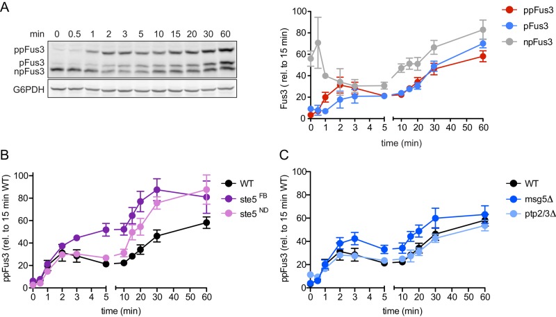
FIGURE 3:
Dynamics of differentially phosphorylated forms of Fus3. (A) Left, wild-type cells were treated for the indicated times with 10 μM α-factor and resolved by Phos-tag SDS–PAGE and immunoblotted with Fus3 antibodies (top) or G6PDH load control antibodies (bottom). Right, dual-phosphorylated (ppFus3), monophosphorylated (pFus3), and nonphosphorylated (npFus3) quantified as a percentage of total Fus3 at 15 min. Results are reported as ±SEM (n ≥ 3). (B and C) Wild-type (WT), ste5FB (feedback-phosphorylation deficient), ste5ND (nondocking), ptp2Δ/ptp3Δ, and msg5Δ (phosphatase-deficient) mutants treated and resolved by Phos-tag SDS–PAGE, as described. Dual-phosphorylated Fus3 is quantified as a percentage of total Fus3 at 15 min. Results are reported as ±SEM (n ≥ 3).

