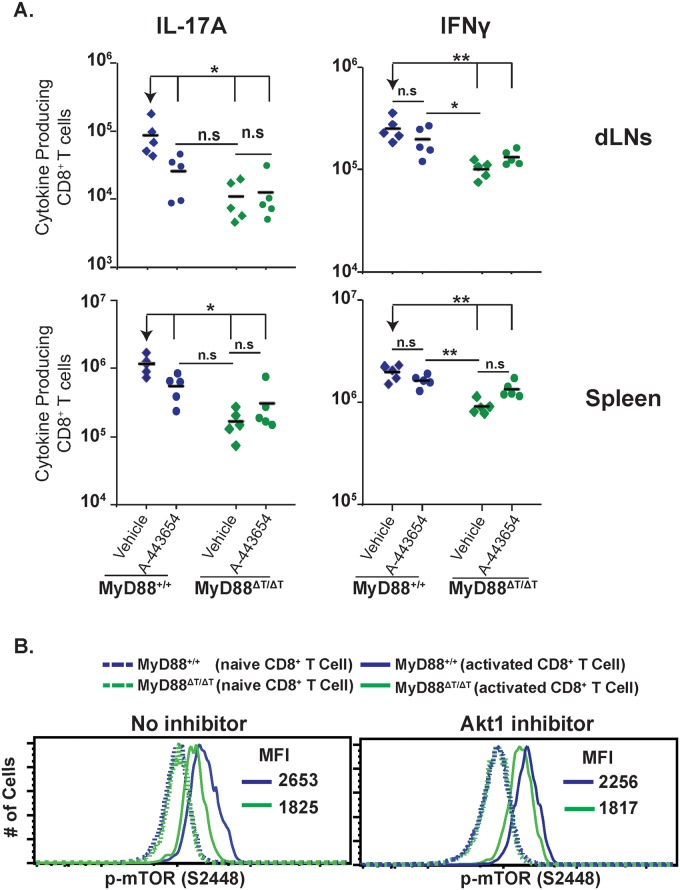
Fig 7. MyD88 requires Akt1 signaling for mTOR activation.
A. Mice were CD4+ T cell depleted and vaccinated with strain #55. Akt1 inhibitor (A-443654) was administered s.c. from day 4 to 15 post-vaccination. On day 16, dLNs and spleen cells were collected, restimulated and stained for surface markers and intracellular cytokines. Numbers of IL-17A and IFNγ producing CD8+ T cells were analyzed by flow cytometry. A diamond represents an individual mouse and the bar is the mean of the group. *p≤0.05 and **p≤0.01. Data is representative of 2 independent experiments. B. Purified naive WT and MyD88ΔT OT-I cells were stimulated in vitro with anti-CD3 and yeast-stimulated BMDC supernatant. On day 4, supernatant was removed and replaced with culture medium. Cells were rested for 2.5 hours and medium was replaced with yeast stimulated BMDC supernatant either with or without Akt1 inhibitor (1μM) for 1 hour. Cells were surface stained prior to phospho-staining. Values in the histograms represent mean fluorescence intensity (MFI) of p-mTOR. Data in solid lines are from gating on activated cells (CD8+CD43+CD44hiCD62Llo), whereas data in dotted lines are from gating on naïve cells (CD8+CD43negCD44loCD62Lhi).

