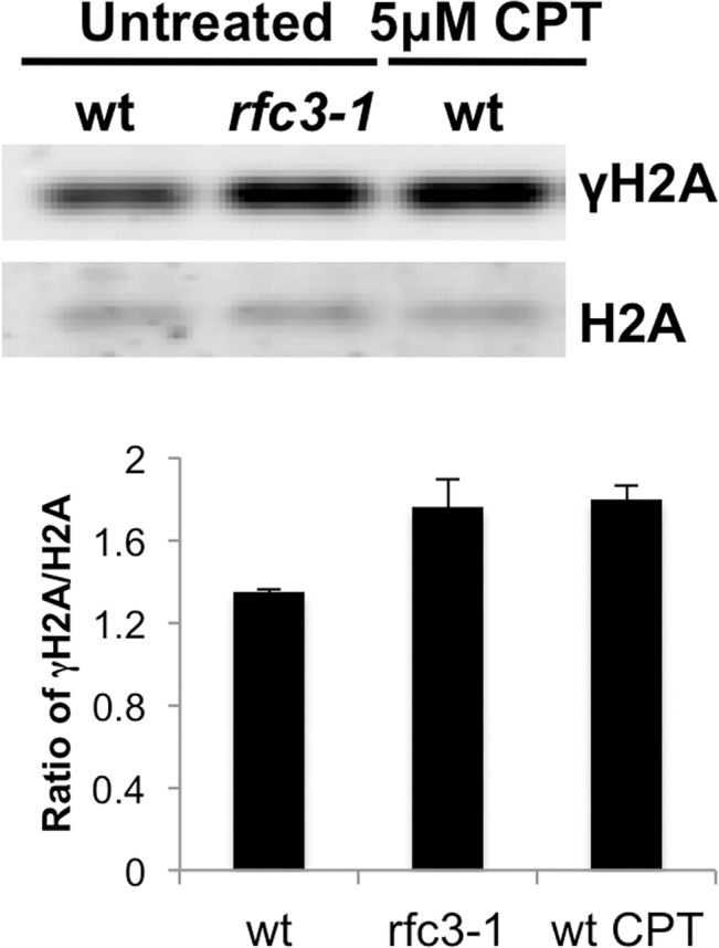
Fig 2. Increased γH2A in rfc3-1 mutant.

Histone enriched cell extracts from the indicated strains were immunoblotted with antisera that bind the C-terminal phospho-SQ epitope of γH2A or H2A itself. Note as shown below and reported previously γH2A in untreated wild type is predominantly from cells passing through S-phase [8]. Note also that rfc3-1 cultures grown at 25°C were previously found to have a DNA content flow cytometry profile similar to wild type [12], indicating that increased γH2A in rfc3-1 cultures most likely arises from increased γH2A-triggering lesions. The increased γH2A in rfc3-1 cells cultured at 25°C is comparable to the level of γH2A in wild type cells treated with 5 μM CPT. Error bars indicate standard error of the mean of 3 independent experiments.
