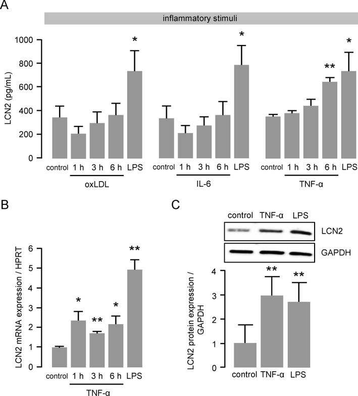
Fig 1. TNF-α enhances LCN2 expression and secretion in macrophages.
(A) LCN2 release from murine primary BMDM following oxLDL (50 μg/mL), IL-6 (200 ng/mL) or TNF-α (50 ng/mL) stimulation was determined by ELISA at the indicated time points. LPS (100 ng/mL for 6 hours) was used as positive control. *P<0.05, **P<0.01 vs. control, n = 6–8 replicated experiments. (B) LCN2 mRNA expression following TNF-α (50 ng/mL) stimulation was determined by real time PCR at the indicated time points. LPS (100 ng/mL for 6 hours) was used as positive control. HPRT was used as housekeeping gene and the relative mRNA expression was calculated using the 2-ΔΔCT method. *P<0.05, **P<0.01 vs. control, n = 4–7 replicated experiments. (C) LCN2 protein expression following TNF-α (50 ng/mL) stimulation for 6 hours was determined by Western blot. LPS was used as positive control. GAPDH was used as loading control. **P<0.01 vs. control, n = 5 replicated experiments.

