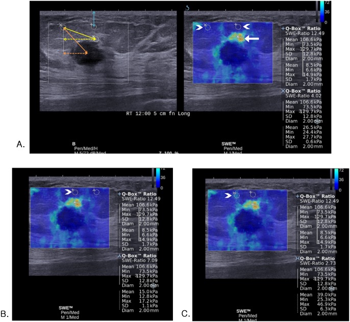
Fig 1. Images of invasive ductal carcinoma in a 54-year-old woman.
(A-C) At a single SWE image obtained from the mass, four Eratios are measured with a fixed ROI for the mass (white arrow) along with four ROIs for the surrounding fat that are set randomly in different four locations (white arrowheads). (A, left) The actual (orange double-headed solid arrow) or vertical (orange double-headed dotted arrow) distance from the center of lesion to the fat ROI, the actual (yellow double-headed solid arrow) or vertical (yellow double-headed dotted arrow) distance from the lesion ROI to the fat ROI, and the vertical distance from the fat ROI to skin (blue double-headed solid arrow) was measured on gray-scale image. (C) For the ROI that is set on the fat tissue showing artifactual vertical light blue color stiffness at SWE (Emean, 39.0 kPa; Emax, 46.9 kPa) (white arrowhead), Eratio was 2.73 which was false negative result according to the cutoff value of 3.18.

