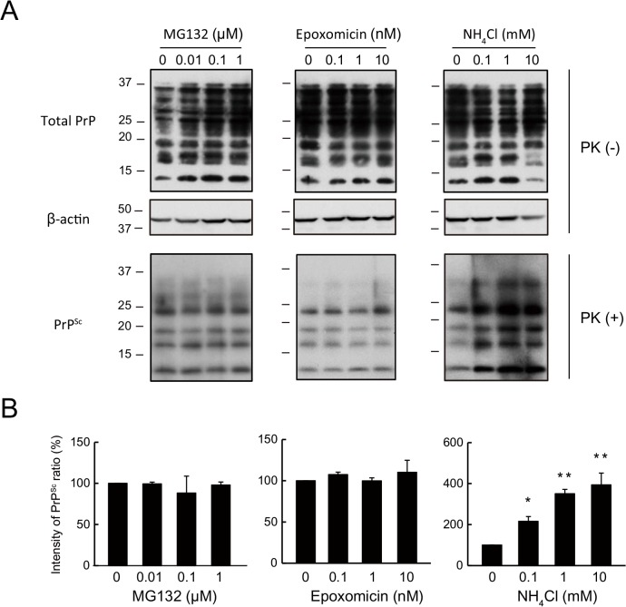
Fig 1. PrPSc in N2a-FK cells is potently increased by a lysosomal but not by a proteasomal inhibitor.
(A) N2aFK cells were treated for 48 h with 0.01 to 1 μM MG132 and 0.1 to 10 nM epoxomicin (Epo) as proteasome inhibitors and 0.1 to 10 mM NH4Cl as a lysosomal inhibitor. PK-treated and-untreated N2a-FK cells were loaded at concentrations of 100 and 30 μg protein per lane onto a 15% polyacrylamide gel and subjected to SDS-PAGE. The proteins were detected by western blotting using anti-PrP and -β-actin antibodies. (B) For densitometric analysis, PrPSc band intensities are expressed as a percentage of those of the negative controls. The results in the graph are the mean ± SD of at least three independent experiments. *p < 0.05 and **p < 0.01 (one-way ANOVA followed by Tukey's test).

