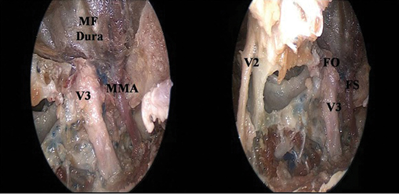
Fig. 4.

Exposure of the infratemporal fossa via the preauricular subtemporal approach (photograph taken with the aid of a 0-degree endoscope [left side]). FO, foramen ovale; FS, foramen spinosum; MF, middle fossa; MMA, middle meningeal artery; V2, maxillary nerve; V3, mandibular nerve.
