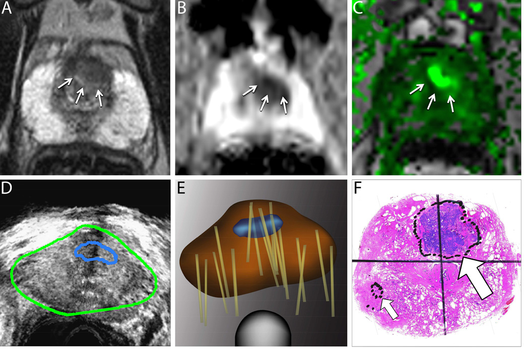
Figure 1. Value of Targeted Biopsy to Evaluate Men for Active Surveillance (Example Case).
A man aged 68 years was referred for active surveillance when a conventional biopsy revealed a small focus of low-grade prostate cancer. A confirmatory biopsy was later performed using magnetic resonance imaging/ultrasound (MRI/US) fusion to target a suspicious region. Top row: Multiparametric MRIs from a T2-weighted image (A), a diffusion-weighted image (B), and a dynamic contrast-enhanced image (C). The delineated region of interest (arrows) was assigned a grade 4 or 5 level of suspicion21. Bottom row: During fusion biopsy, the region of interest from an MRI is superimposed on a US image (D). Systematic and targeted biopsy cores (tan lines) were obtained and recorded on a digital reconstruction of the prostate using an MRI/US fusion device (Artemis; Eigen, Grass Valley, Calif) (E).
A targeted biopsy revealed potentially lethal, high-grade cancer, and radical prostatectomy was performed. A whole-mount prostate sample reveals high-grade cancer (large arrow) that was not detected on an earlier conventional biopsy (F). The small arrow indicates a focus of low-grade cancer that was identified on an earlier “blind” biopsy. Targeted biopsy provides more accurate characterization of whole-gland pathology than conventional (blind) biopsy and is increasingly used to evaluate men for active surveillance34. In the example case, a targeted biopsy revealed that the patient was a poor candidate for active surveillance. Reprinted with permission, American Cancer Society47.

