
Figure 1. Schematic of oligodendrogenesis after demyelination.
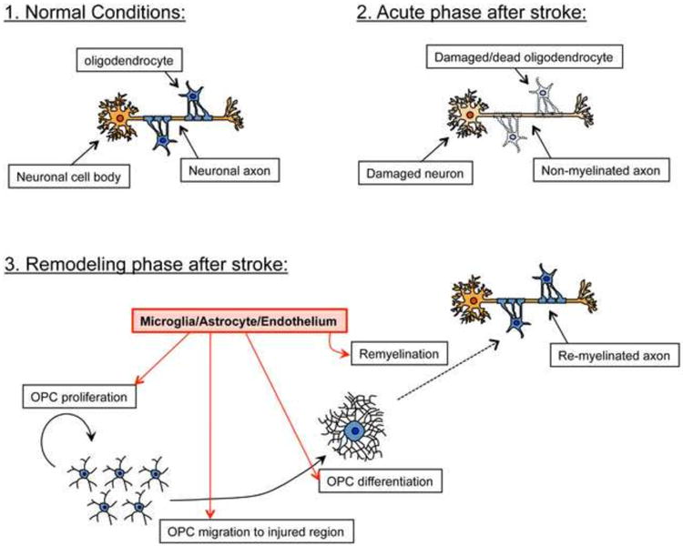
Under normal conditions, neuronal axons receive support from myelinated oligodendrocytes. After injury such as stroke, damaged oligodendrocytes no longer support axons, resulting in failure of proper neuronal function. However, during the remodeling phase after stroke, residual OPCs (or OPCs differentiated from NSPCs) proliferate, migrate to the injured region, and then differentiate into oligodendrocytes which help to restore the damaged myelin sheaths. Importantly, neighboring cells including microglia, astrocytes, cerebral endothelial cells, also actively support oligodendrogenesis by secreting soluble trophic factors. It is unclear, however, whether neuronal axons that have been damaged after stoke, and subsequently repaired through the process of oligodendrogenesis, may be able to regain normal myelin function.